 In June 2022, a team led by Dr. Yueyi Yu from Capital Medical University Beijing (China) published an article in Frontiers in Neuroscience reporting that they found acanthocytes in the blood of people with Huntington's disease (HD). While they only found these cells in four out of 40 people with HD, the authors concluded that acanthocytosis might be a new clinical characteristic of HD.
Thorny, misshapen red blood cells, referred to as “acanthocytes”, are found in several diseases, such as chorea-acanthocytosis (VPS13A disease) and McLeod syndrome (XK disease). They can also be seen with severe liver problems, malnutrition or vitamin deficiency. However, acanthocytosis (the presence of these misshapen red blood cells) has been always regarded as a feature which distinguishes HD from the so-called “neuroacanthocytosis syndromes” (VPS13A disease and XK disease).
For this reason, our community took note of this article. After thoroughly studying the manuscript and figures we concluded that the majority of misshapen red blood cells shown in this work should be classified as “echinocytes” rather than acanthocytes. Echinocytes are less misshapen, and have more regular spines on their surfaces, and can occur with various forms of red blood cell (physical) stress and do not indicate any particular disease.
Therefore, we strongly doubt that the conclusion from the Beijing group is warranted, and we submitted a commentary article on this issue to Frontiers in Neuroscience. Here, we further explained the reasons for our judgement and extended the discussion on other related topics, such as the classification of misshapen red blood cells and the difficult role of acanthocyte testing for clinical diagnosis in general. We also proposed new methods for the exact determination of red blood cell shapes using artificial intelligence. In addition, we suggested that the diagnosis of “neuroacanthocytosis” should not rely solely on the presence or absence of acanthocytes, but should instead include a broader spectrum of laboratory tests.
Further studies are needed to determine if misshapen red blood cells actually occur in HD patients. This will extend our understanding of similarities and differences between HD and neuroacanthocytosis syndromes.
For further reading, please refer to:
Peikert K, Storch A, Hermann A, Landwehrmeyer GB, Walker RH, Simionato G, Kaestner L, Danek A. Commentary: Acanthocytes identified in Huntington's disease. Front Neurosci. 2022 Nov 4;16:1049676. doi: 10.3389/fnins.2022.1049676.
https://www.frontiersin.org/articles/10.3389/fnins.2022.1049676/full
Original article describing acanthocytes in Huntington’s disease patients:
Yu Y, Lu Y, Wang F, Lu Y, Xie B, Meng X, Tang Y. Acanthocytes Identified in Huntington's Disease. Front Neurosci. 2022 Jun 6;16:913401. doi: 10.3389/fnins.2022.913401
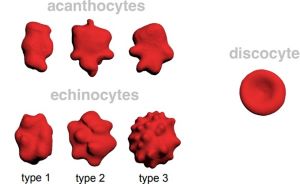
Figure legend
Distinction of acanthocytes from echinocytes based on 3D images and classification with artificial neural networks as proposed by our group (figure modified from Peikert K, Storch A, Hermann A, Landwehrmeyer GB, Walker RH, Simionato G, Kaestner L, Danek A. Commentary: Acanthocytes identified in Huntington's disease. Front Neurosci. 2022 Nov 4;16:1049676).
Discocytes are normal red blood cells. There are many different types of malformed red blood cells. Thorny, irregularly misshapen red blood cells are referred to as acanthocytes, whereas cells with many small, evenly distributed thorny projections are called echinocytes.]
https://www.frontiersin.org/articles/10.3389/fnins.2022.913401/full

 Like
Like