 Adrian Danek and Gabriel Miltenberger-Miltenyi in Munich
The working group of Adrian Danek and Gabriel Miltenberger-Miltenyi in Munich continue with the free-of-charge chorein blot diagnosis, supported by the generous donation of seed money by Betty and Carl Pforzheimer to the Advocacy, in honour and in memory of Glenn Irvine.
Reports containing the obtained results for each patient are sent to the submitter and a detailed discussion of every case is offered.
In parallel, the Munich team continues with the genetic testing of ChAc patients; for details please contact danek@lmu.de or gmiltenyi@medicina.ulisboa.pt.
Currently, one of the major technical-scientific questions the team is focusing on, is the occurrence of rare cases of patients with seemingly normal chorein band occurrence in the Western blot, despite pathogenic mutations in the VPS13A gene. Six such cases were detected so far. This challenging question is supposed to be solved by genotype-phenotype association studies.
In addition, the team in Munich continues with the development of the zebrafish model for ChAc and McLeod Syndrome in collaboration with local colleagues at the German Center for Neurodegenerative Diseases (DZNE).
PHOTO: Gabriel Miltenberger-Miltenyi (L) and Adrian Danek (R)
_____
Targeting Lyn kinase in chorea-acanthocytosis: First experiences with a potentially disease modifying approach
Kevin Peikert, Andreas Hermann and Lucia De Franceschi
 In red blood cells and neurons of chorea-acanthocytosis (ChAc) patients, the enzyme “Lyn kinase” is overactive which leads to impairment of some cellular processes. Lyn kinase is therefore considered to be one of the drivers of the disease. Suppression of its activity by several compounds can partially restore these damages in cell culture models. Fortunately, some of these drugs with the potential to inhibit Lyn are already in clinical use for leukaemia patients.
Based on their preclinical work with Lyn kinase, an international consortium led by Andreas Hermann (University Rostock, Germany) and Lucia De Franceschi (University of Verona, Italy) sought to “translate” this knowledge as soon as possible to the benefit of the patients into the clinic.
First three patients were treated with an FDA approved Lyn kinase inhibitor, dasatinib. It was promising to see that some blood parameters responded to the treatment, however, there was no clinically evident positive effect on the central nervous system, e .g. no changes in seizure frequency or movement disorder. During these individual treatments, it became also clear that we do not know enough about the natural history of ChAc and about markers that reflect the progress of the disease – so called biomarkers. Altogether, it makes it difficult to judge if an intervention is effective or not, especially in such a small group of patients.
 To further investigate the effect of Lyn kinase inhibition, we used a mouse model of ChAc. We first investigated the role of Lyn in basal ganglia from ChAc mice and then we tested dasatinib and nilotinib. In ChAc mice, it became clear that dasatinib did not sufficiently reach the most important part of the brain for this disease, the basal ganglia, which could be the reason for the lacking effect in the central nervous system of the individual patients. However, a substance of a next generation, nilotinib, was able to penetrate this region and to ameliorate several disease characteristics in this brain region.
At this point, the consortium gathered some promising data on a potentially beneficial effect of Lyn kinase inhibition for ChAc patients. However, the evidence is clearly still too preliminary to generally recommend this treatment approach in ChAc. We will keep the Neuroacanthocytosis community informed as soon as we have further evidence on the efficacy of this therapy.
We thank the Advocacy for Neuroacanthocytosis patients for the ongoing support and the provided funding. We thank also neuroacanthocytosis patients and their families for keeping high the attention on this rare disease and for being part of our lives as scientists.
TOP PHOTO: Andreas Hermann (L) and Kevin Peikert (R)
2ND PHOTO: Lucia De Franceschi
The studies have been published here:
Therapeutic targeting of Lyn kinase to treat chorea-acanthocytosis
Peikert K*, Federti E*, Matte A, Constantin G, Pietronigro EC, Fabene PF, Defilippi P, Turco E, Del Gallo F, Pucci P, Amoresano A, Illiano A, Cozzolino F, Monti M, Garello F, Terreno E, Alper SL, Glaß H, Pelzl L, Akgün K, Ziemssen T, Ordemann R, Lang F, Brunati AM, Tibaldi E, Andolfo I, Iolascon A, Bertini G, Buffelli M, Zancanaro C, Lorenzetto E, Siciliano A, Bonifacio M, Danek A, Walker RH, Hermann A*, De Franceschi L*. May 2021. Acta Neuropathol Commun 9:81. doi:10.1186/s40478-021-01181-y. *contributed equally.
Targeting Lyn Kinase in Chorea-Acanthocytosis: A Translational Treatment Approach in a Rare Disease
Peikert K, Glaß H, Federti E, Matte A, Pelzl L, Akgün K, Ziemssen T, Ordemann R, Lang F, The Network for Translational Research for Neuroacanthocytosis Patients, De Franceschi L*, Hermann A*. May 2021. Journal of Personalized Medicine 11(5):392. . *contributed equally.
_____
The Erythrocyte Sedimentation Rate may serve as a novel biomarker for Neuroacanthocytosis Syndromes
Kevin Peikert & Lars Kaestner
 The erythrocyte sedimentation rate (ESR) is one of the oldest diagnostic methods. Already in ancient times it was known that the sedimentation of the red part of the blood can be very different. In fact, it was known even before the thermometer was invented that many diseases cause a higher ESR, which as we know today is also related to increased body temperature.
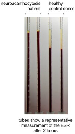
Medically, there are two effects involved: (i) the increase of plasma proteins associated with inflammation, and (ii) also the erythrocyte shape contributes to the ESR. Irregular cell shapes, like sickle cells or acanthocytes decrease the ESR. So far in medical practice, no lower limit was established as being pathologic.
Recently, in a collaborative project between the neurologists at the University Hospitals in Rostock (Kevin Peikert, Hannes Glass and Andreas Hermann) and Munich (Adrian Danek) with biophysicists at Saarland University (Alexis Darras and Lars Kaestner, all Germany), a low ESR was proposed as a novel biomarker for the neuroacanthocytosis syndromes chorea-acanthocytosis and McLeod syndrome (Fig. 1). The clinicians and scientists summarized their results in two articles recently published in scientific journals: (Darras et al. 2021; Rabe et al. 2021).
 Although a lower limit for the ESR was found, these are just initial results that require further studies with a higher number of volunteers to proof the methods specificity. Furthermore, it remains still elusive if the ESR can be used as a putative biomarker for diagnosis, for disease progression of individual patients or response to a potential treatment. However, on the basis of these new data, the authors recommend measuring the ESR in all patients with suspected neuroacanthocytosis syndromes.
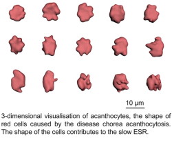
Beside the fact that the ESR is among the oldest diagnostic methods, these measurements were combined with the latest scientific advancements in optical 3-dimensional microscopy and the application of artificial neuronal networks to analyze the data (Fig. 2).
Independent of the neuroacanthocytosis syndrome, the ESR was physically described in a completely new manner. Historically, ESR was described as sedimenting aggregates of red cells, but now it can be better understood as a percolating gel formed by the red cells in stasis.
Figure 1 (R) Figure 2 (L)
|
|
|
TOP PHOTO: Kevin Peikert
2ND PHOTO: Lars Kaestner
The studies have been published here:
Darras, Peikert, Rabe, et al (2021) Acanthocyte sedimentation rate as a diagnostic biomarker for neuroacanthocytosis syndromes: experimental evidence and physical justification. Cells 10:788.
Rabe A, Kihm A, Darras A, et al (2021) The Erythrocyte Sedimentation Rate and Its Relation to Cell Shape and Rigidity of Red Blood Cells from Chorea-Acanthocytosis Patients in an Off-Label Treatment with Dasatinib. Biomol 11:727.
_____
“Neuroacanthocytosis” – Overdue for a Taxonomic Update
Ruth Walker & Adrian Danek
 The term 'neuroacanthocytosis' (NA) is used for a spectrum of neurological disorders in which there are thorny red blood cells. While NA historically referred to disorders of lipoprotein absorption, we have promoted it as an overarching term for a group of basal ganglia disorders, with specific reference to two diseases that we defined as 'core' NA syndromes. 'Neuroacanthocytosis' has also been used to refer to a specific, now genetically-defined disease, otherwise known as 'chorea-acanthocytosis'. These various usages have resulted in diagnostic confusion, and in a number of cases have quite likely prevented the pursuance of precise, molecular, diagnosis. Disease nomenclature is an everevolving field, especially in the current era of expanding genetics, and naming proposals are often far from ideal. We, however, suggest that the term 'neuroacanthocytosis' should no longer be generally used and if so, only with appropriate understanding of its limitations. Further, we propose that chorea-acanthocytosis be renamed as “VPS13A disease” in accordance with its genetic etiology.
PHOTO: Ruth Walker (L) and Adrian Danek (R)
The study has been published here:
Walker RH, Danek A. “Neuroacanthocytosis” – Overdue for a Taxonomic Update. Tremor and Other Hyperkinetic Movements. 2021;11(1):1. DOI:
_____

 Like
Like